Phone: (714) 695-1566
Fax: (714) 695-1553
Email: info@salinaspt.com
23655 Via Del Rio, Suite C
Yorba Linda, CA 92887
Phone: (714) 695-1566
Fax: (714) 695-1553
Email: info@salinaspt.com
23655 Via Del Rio, Suite C
Yorba Linda, CA 92887

The syndesmotic or high ankle sprain typically occurs during a forceful turning in or out of the lower leg when your foot is flexed upward as seen above. It almost always happens during a collision, not by simply rolling your ankle.
When Patrick Mahomes was tackled, the defensive player landed on his leg, causing the inside of the foot to roll outward toward the small toes. This motion stressed the high ligaments known as the interosseus membrane in his leg.
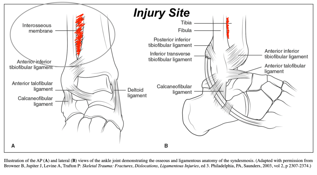
Red contrast added to indicate injury location.
A thorough history can often provide clues when questioning the mechanism of injury. The physical exam includes several special tests, including the squeeze, external rotation stress test, fibular rotation, and cotton tests. The squeeze test is done by the practitioner squeezing the tibia and fibula together, stressing the interosseus membrane. While seated with legs dangling, pushing up on foot and rotating the tibia outward can also evaluate the membrane and help diagnose this injury. Imaging studies are also performed to assess the extent of the damage.
Several classification systems can be used to classify the type of injury. Most ankle classification systems use severity as a way to categorize ligament injuries.
Most high ankle sprains are treated conservatively (non-operative) with ankle splinting to reduce the painful joint’s motion. Surgery is rarely necessary for high ankle sprains. It is indicated only if a significant injury to the ligaments around the ankle results in visable separation of the tibia and fibula. During the acute phase, protection, rest, ice, compression, and elevation (PRICE) will control pain and swelling. Crutches are sometimes used to limit weight bearing. Often the ankle will be placed in a walking boot to allow adequate healing.
Once the acute inflammatory phase is over and the pain and swelling have begun to subside, gentle range of motion exercises are started as tolerated. This is followed by the first phase of strengthening exercises for the lower leg muscles. Mobility exercises are initiated to regain a normal walking pattern and resume normal activity levels.
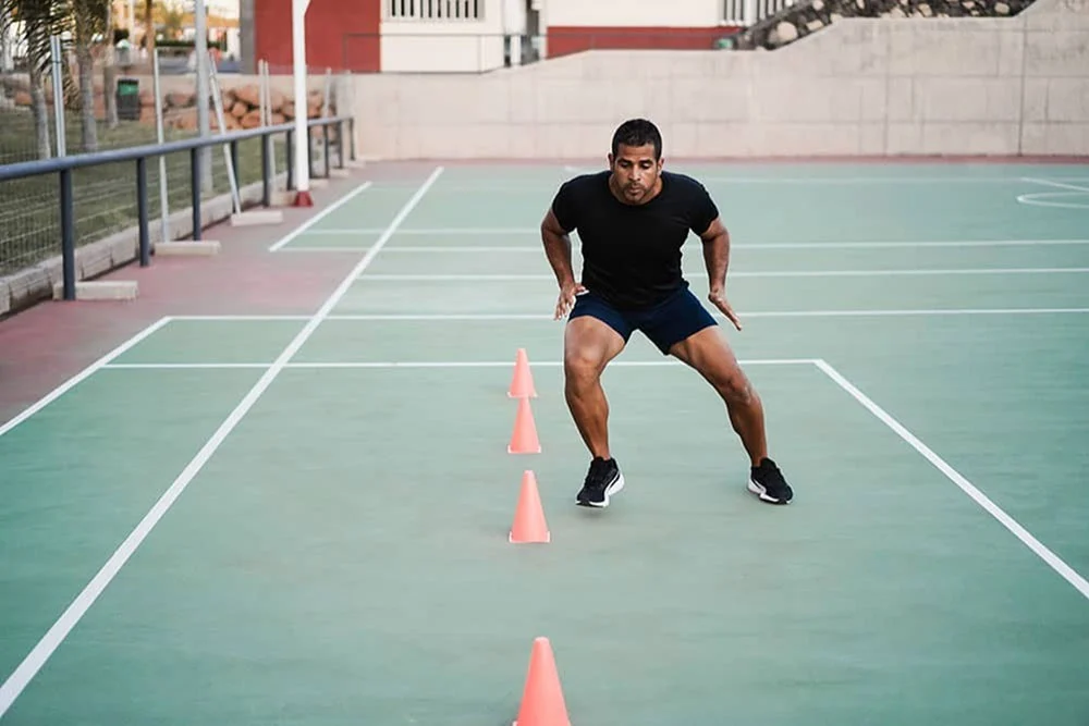
As the strength and stability of the injured ankle return to normal, the progression of sport-specific exercises is then initiated to prepare the athlete to return to sports. Specific criteria must be met for the athlete to compete safely. The ability to run, cut, accelerate, and decelerate must all be restored.
What’s most remarkable about Patrick Mahomes’ recovery was that he first injured his right ankle against the Jacksonville Jaquars on January 21. He returned that game in the second half to help the Chiefs win 27-20. In a matter of weeks he was not only able to compete, but take home Lombardi trophy post-injury. The Kansas City Chiefs sports medicine staff managed his injury remarkably and prepared him for one of the biggest games of his life.
We understand that suffering from an injury can take you out of your “game”. From walking the dog, to trimming the garden hedges, we all rely on our physical ability to support the lifestyles we live, or want to live. One of the key takeaways of this message is that your body is an amazing structure and it should be treated that way. By implementing healthy lifestyle decisions you can expect positive outcomes well into your golden years. If you’re looking for more ways improve your health, check out the Healthy Living section on our Blog or contact us using the link below. nnn